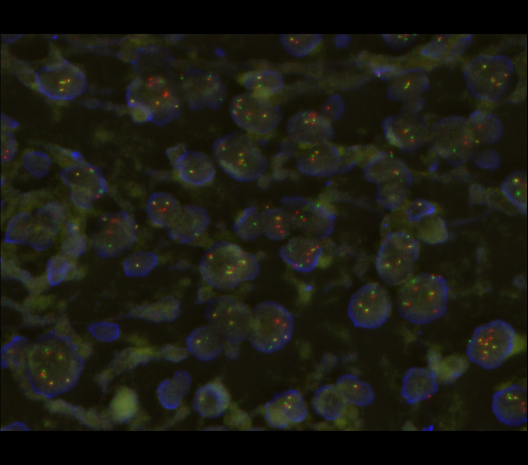
Figure 3.

Fluorescence in situ hybridization (FISH) micrograph, demonstrating rearrangements of the 5’ and 3’ regions of the EWSR1 gene labeled in red and green, respectively, with occasional yellow signals denoting intact EWSR1 genes. Nuclear DNA is labeled in blue.
