Fig. 1.
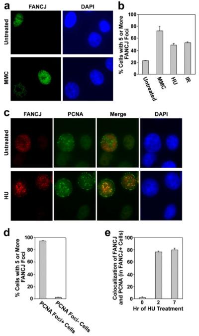
FANCJ nuclear foci assemble spontaneously in S phase cells, are induced by various stresses, and are recruited to blocked replication forks. a Examples of FANCJ nuclear foci (green) in untreated populations of MCF7 cells or following treatment of cells with 0.5 μM MMC for 20 h. Nuclei were stained with DAPI (blue) for visualization. b Quantification of the percentage of MCF7 cells that displayed five or more FANCJ foci in untreated populations, following exposure to 0.5 μM MMC or 2 mM HU for 7 h, or at 7 h following exposure to 5 Gy IR. c Examples of FANCJ nuclear foci (red) and PCNA foci (green) in untreated MCF7 cells or following treatment with 2 mM HU for 20 h. The degree of colocalization is demonstrated in the merged image. d The percentage of S phase or non-S phase cells, identified by the presence or absence of nuclear PCNA signal, respectively, that displayed five or more FANCJ foci. e The percentage of cells with five or more FANCJ nuclear foci that displayed colocalization with PCNA foci in untreated MCF7 cells, or at 2 or 7 h of treatment with 2 mM HU. PCNA is a marker for the replication fork. Quantitative values represent the average of three counts of 150 or more cells each±standard deviation (b, d–e). The levels of FANCJ foci, or colocalization of FANCJ and PCNA foci, were significantly increased (p<0.01) following treatment with HU or DNA damage in b and e, respectively

