Fig. 3.
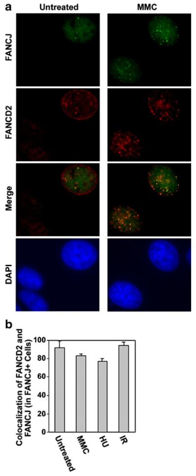
FANCJ and FANCD2 foci partially colocalize in untreated MCF7 cells and following exposure to various types of DNA damage. a Examples in which FANCJ (green) and FANCD2 nuclear foci (red) colocalized in untreated populations of MCF7 cells or following treatment of cells with 0.5 μM MMC for 20 h. b Quantification of the percentage of MCF7 cells with FANCJ foci that displayed three or more colocalized FANCD2 foci in untreated populations, following exposure to 0.5 μM MMC or 2 mM HU for 7 h, or at 7 h following exposure to 5 Gy IR. Results represent the average of three counts of 150 or more cells each±standard deviation

