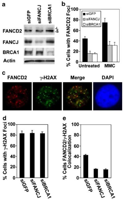
Fig. 6.
FANCJ is involved in the recruitment of FANCD2 to DNA double-strand breaks. a Immunoblots to detect FANCD2, FANCJ, or BRCA1 in extracts from untreated MCF7 cells transfected with a control siRNA directed against GFP, or with siRNAs against either FANCJ or BRCA1. Both unubiquitinated (-S) and monoubiquitinated (-L) FANCD2 were detected with E35 antibody. Actin is shown as a loading control. b Quantification of the assembly of FANCD2 nuclear foci in MCF7 cells transfected with siGFP (control), or with siRNAs targeting FANCJ or BRCA1. Cells were left untreated or were incubated with 0.5 μM MMC for 20 h. c An example of the colocalization of FANCD2 foci (red) with DNA double-strand breaks, detected with an antibody against γ-H2AX (green), in MCF7 cells at 7 h following exposure to 5 Gy IR. Colocalization is evident in the merged image. Nuclei were identified by staining with DAPI. d Quantification of the percentage of MCF7 cells transfected with the indicated siRNAs that displayed γ-H2AX foci at 7 h after exposure to 5 Gy IR. e Quantification of the percentage of MCF7 cells, transfected with indicated siRNAs which displayed colocalization of five or more FANCD2 and γ-H2AX foci after exposure to IR. Average values were calculated from three counts of 150 or more cells each and are displayed±standard deviation (b, e). The levels of FANCD2 foci and its colocalization with γ-H2AX foci were statistically different (p<0.01) in cells depleted of FANCJ or BRCA1, as compared with cells transfected with the control siRNA (b, e)

