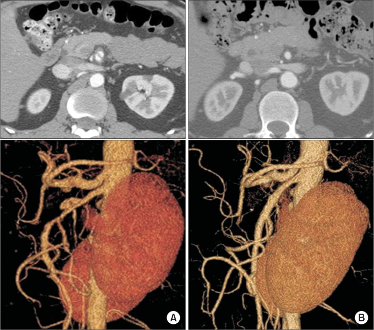
Fig. 5.
Complete remodeling of superior mesenteric artery (SMA) dissection on follow-up computed tomography (CT) angiogram. (A) Initial CT scan showing double-lumen sign on an axial view (top) and a wind-sock-shaped dissection lesion on the anterior wall of the SMA (type IIa spontaneous isolated superior mesenteric artery dissection) (bottom), (B) follow-up CT angiogram at 7 months after conservative treatment shows disappearance of the double-lumen sign on axial view (top) and complete remodeling of the SMA on a reconstructed view of the SMA (bottom).

