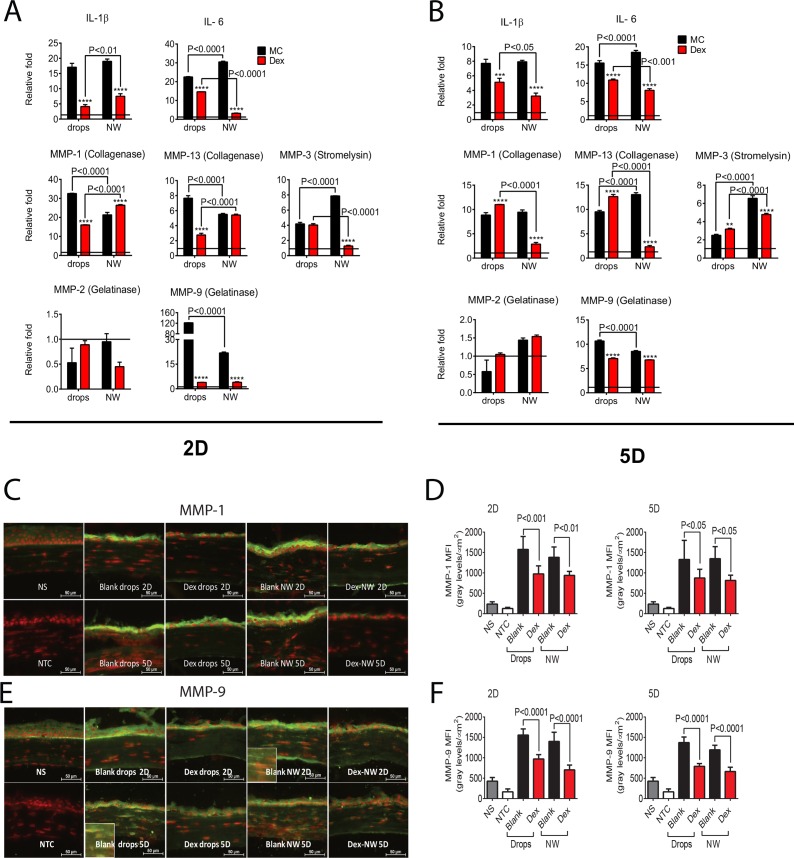
Figure 3.
Methylcellulose NW loaded with 10 μg Dex decrease inflammatory cytokines and MMPs in corneas of the combined model of alkali burn and dry eye. (A, B) Mean ± SEM of results of gene expression analysis of inflammatory cytokines (IL-1β and IL-6) and MMPs-1, -2, -3, -9, and -13 RNA transcripts in whole corneas from animals subjected to ocular burn + desiccating stress for 2 (A) or 5 (B) days and topically treated with Dex drops or Dex-NW and compared with its vehicle controls. The horizontal line at each figure represents the level of the mRNA expression for untreated group, which was used as the calibrator and normalized as 1. n = 4–5 right corneas/group. (C, D) Representative merged pictures of MMP-1 (C) and -9 (D) immunofluorescent staining shown in green of central cornea cryosections from animals subjected to a combined model of alkali burn and dry eye topically treated with Dex drops or Dex-NW and compared with its vehicle controls. Counterstaining was PI = red; n = 6 right corneas/group. (E, F) Quantification of mean fluorescence intensity (MFI) values from MMP-1 (E) and MMP-9 (E) staining are displayed. n = 6 right corneas/group. n = 4–5 right corneas/group. *P < 0.05, **P < 0.01, ***P < 0.001, ****P < 0.0001: MC blank wafers versus MC Dex-NW, MC blank drops versus MC Dex drops. MC blank-NW versus MC Dex-NW; NS = nonstressed; NTC, negative control; MFI, mean fluorescence intensity.

