Figure 1.
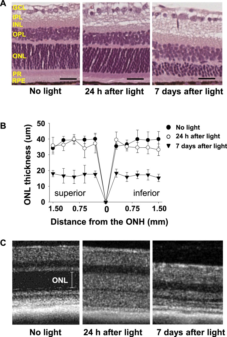
Retinal changes after light exposure in Abca4−/−Rdh8−/− mice. (A) Representative retinal images of the inferior retina at 750 to 1000 μm from the optic nerve head (ONH) of 4-week-old Abca4−/−Rdh8−/− mice after light exposure at 10,000 lux for 30 minutes are shown. No apparent morphologic alterations are visible 24 hours after light exposure whereas severe retinal degeneration is observed 7 days after light exposure (n = 3 for each group). PR, photoreceptors; OPL, outer plexiform layer; INL, inner nuclear layer; IPL, inner plexiform layer; GCL, ganglion cell layer. Scale bar: 20 μm. (B) Outer nuclear layer thickness of 4-week-old Abca4−/−Rdh8−/− mice after light exposure at 10,000 lux for 30 minutes is plotted (n = 3 for each group). Sections were prepared with the paraffin embedment. Error bars: Mean ± SD. (C) Representative in vivo SD-OCT imaging of the inferior retina at 500 to 750 μm from the ONH show changes in ONL thickness 24 hours after light exposure and by 7 days there is an observed loss of ONL indicating severe retinal degeneration (n = 3 for each group).

