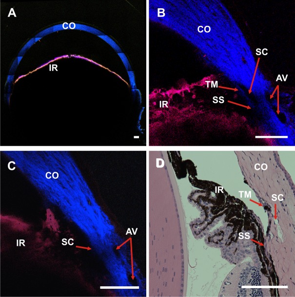
Figure 1.
Spectral TPM of TPAF and SHG images of an unfixed, enucleated C57BL/6 mouse eye. (A) A TPM composite image tiled from a 7 × 7 image array shows the entire anterior cross-section (pixel size: 0.692 μm). Red: TPAF; blue: SHG. (B) and (C) are single TPM images of the limbal region from two different C57BL/6 mice (pixel size: 0.259 μm). The IR is visible by the TPAF signal (red) of the tissue melanin, and the collagen from the CO generates strong SHG (blue). Aqueous veins are identified by the absence of the SHG signal near the corneal/scleral junction. Schlemm's canal is identifiable as an area lacking SHG signal near the insertion point of the IR. The TM layer could be identified in (B) as a thin strip of faint SHG signal immediately adjacent to the SC. (D) A hematoxylin and eosin–stained histologic section of a C57Bl/6 eye. The relative locations of the SC, TM, and IR are in close proximity to those identified in the TPM images. SS, scleral spur. Scale bar: 100 μm.

