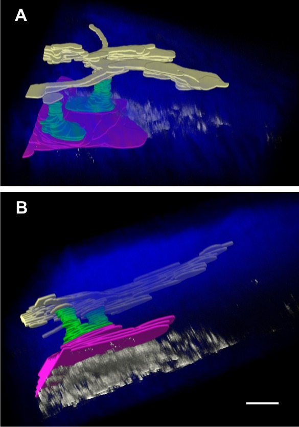
Figure 6.

Three-dimensional image reconstruction and segmentation of the AOS of a C57BL/6 mouse eye perfused with 0.1 μm polystyrene beads through the cornea into the anterior chamber. (A) View from outside the eye looking into sclera, and (B) the same reconstruction viewed from the side. The fluorescent beads (white) accumulate beneath the SC (red) where the entrances of CCs (green) were located. There were a few fluorescent beads identified within the SC, CCs, and AVs (yellow). Scale bar: 30 μm in the x- and y-axes and 15 μm in the z-axis (the z-axis is dilated by a factor of 2).
