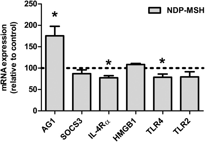
Fig 1. Effect of NDP-MSH on the expression of M2 markers and inflammatory mediators.
Cells were treated with 100 nM NDP-MSH for 24 hours. Gene expression was assessed by RT-qPCR. Values are the mean ± SEM of at least 3 independent experiments and are expressed as the percentage of their respective controls (arbitrarily set at 100% and represented by the dotted line). Data were analysed by one-sample t test. *p<0.05 vs. control group.

