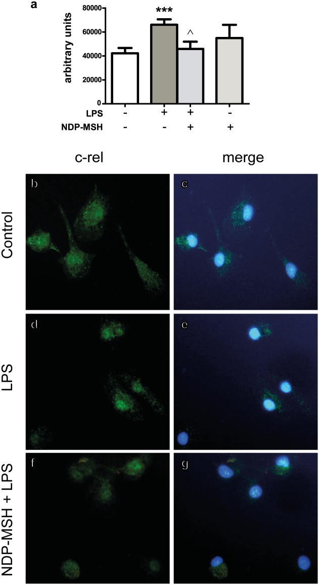
Fig 3. NDP-MSH prevents LPS-induced c-Rel nuclear translocation.
Cells were treated for 24 hours with 100 nM NDP-MSH and then stimulated for 30 minutes with LPS (100 ng/ml) or Pam3CSK4 (100 ng/ml) and processed for c-Rel immunocytochemistry. (a) Nuclear fluorescence intensity was semi-quantified using ImageJ software. Data are the mean ± SEM of 4 independent experiments and were analysed by one-way ANOVA followed by Bonferroni’s multiple comparisons test. ***p<0.001 vs. control. ^p<0.01 vs. LPS. Representative images are shown of the following groups: (b) and (c) Control; (d) and (e) LPS; (f) and (g) NDP-MSH + LPS. Green: c-Rel. Blue: nuclei stained with DAPI.

