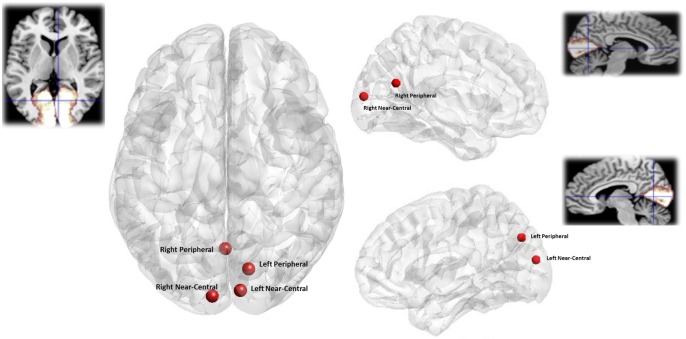
Fig 2. Location of visual stimuli responses.
Visual cortex responses to each stimulus type averaged across all participants a) Axial view b) Left Hemisphere (sagittal view) c) Right Hemisphere (sagittal View). Insets show an anatomical mask of the primary visual cortex (BA17) derived from the Anatomy Toolbox http://www.fz-juelich.de/. All locations are based on MNI co-ordinates.

