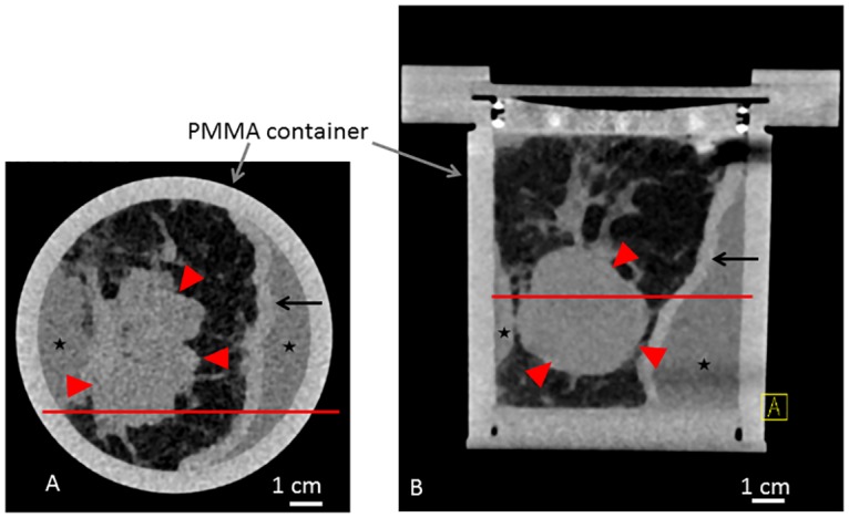
Fig 2. Clinical CT scan of sample A.

Clinical CT scan of sample A in the sample holder with a standard clinical whole body CT scanner (Somatom Definition Flash, Siemens, Germany). Axial (A) and coronal (B) view. Red line in (A) and (B) reference for the respective imaging position. Asterisk indicating formaldehyde surrounding the sample. Red arrowheads indicating tumor. Arrows indicating skin.
