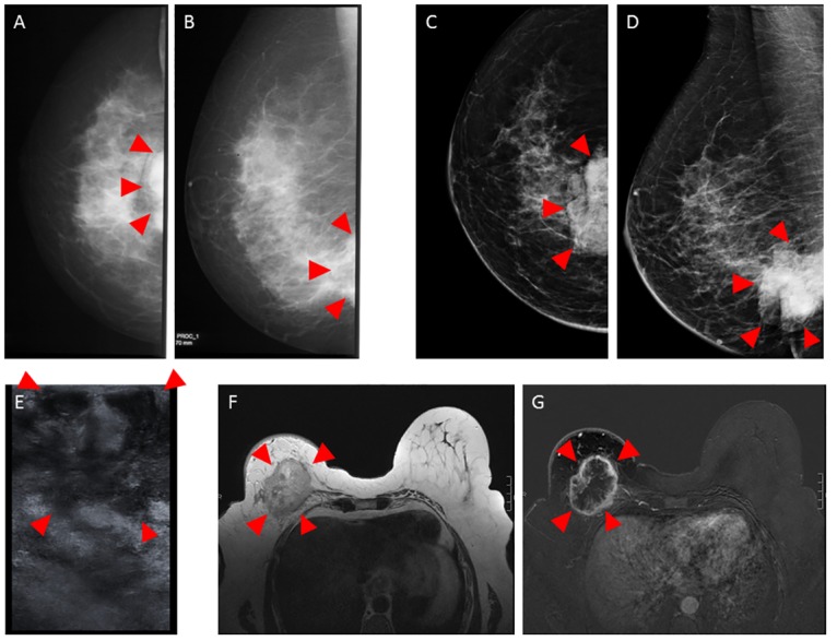
Fig 7. Clinical mammography, sonography and MRI of patient A.

In-vivo mammography of patient A in craniocaudal (A and C) and mediolateral oblique (B and D) projection before (A and B) and after completion of NAC (C and D). Ultrasound before NAC (E). In-vivo MRI including contrast-enhanced T1 weighted gradient-echo sequence after manual injection of 30 ml gadopentetate dimeglumine (Magnevist ® 0.5 mmol/ml) (F) and the corresponding first subtraction image after 2 min (G) in an axial view using a dedicated sensitivity-encoding enabled bilateral breast coil with a 1.5-Tesla system. The tumor is marked with arrowheads. Please note that the tumor is only partially imaged in conventional mammography due to the prepectoral position (A–D) and in Ultrasound (E) due to the extensive size.
