
Fig 8. Clinical mammography and sonography of patient B.
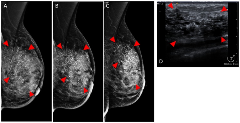
In-vivo mammography in mediolateral oblique projection before (A), after 4 cycles NAC (B) and after completion of NAC (C). Ultrasound before NAC (D). Tumor indicated by arrowheads. Please note the excessive calcifications (white spots in A–D) within the tumor. The exact tumor borders are not visible in mammography and sonography.