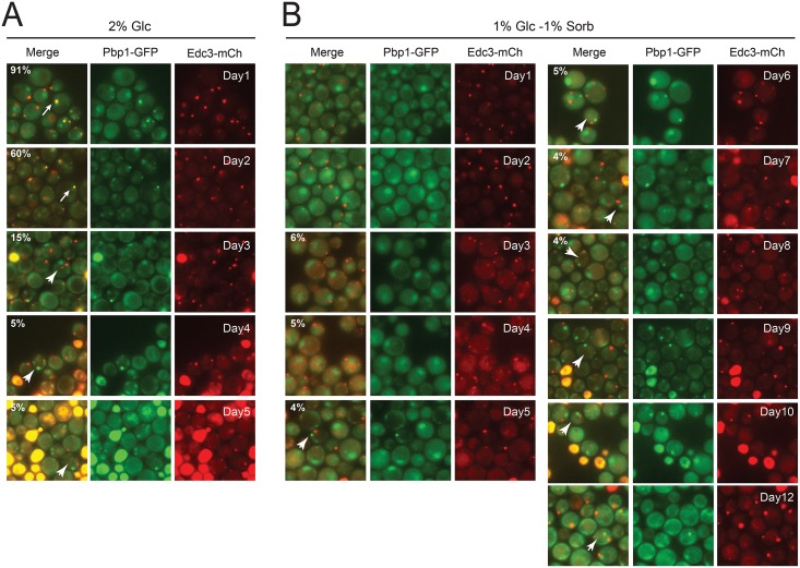
Fig 4. Hybrid-bodies were present only transiently in cells.
Wild-type cells were grown in SC minimal media containing either 2% glucose or a mixture of 1% glucose and 1% sorbitol for the indicated number of days before being examined by fluorescence microscopy. The numbers in the top left corner of the merged image panels indicate the relative level of colocalization observed for the stress granule (Pbp1-GFP) and P-body (Edc3-mCh) reporters present. The white arrows point out cells with colocalized reporters (containing hybrid-bodies) whereas the white arrowheads indicate cells with separate P-body and stress granule foci. The cells grown in the medium containing 2% glucose exhibited a significant level of cell death with increasing time and thus were examined only during the first five days of culture growth.

