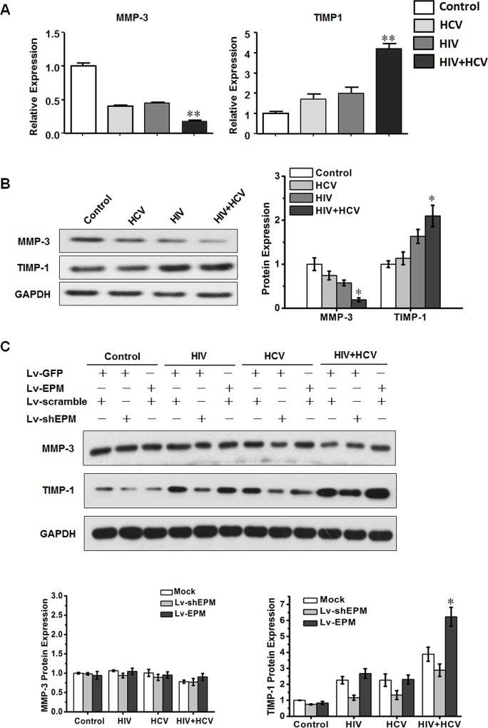
Fig 4. HIV+HCV co-culture increased tissue inhibitors of MMP 1 (TIMP-1) expression levels via an EPM dependent signaling pathway.
(A) QRT-PCR analysis of TIMP-1 and MMP-3 expression levels in LX-2 cells incubated with control medium, HCV (JFH1), inactivated HIV (NL4-3) or HIV and HCV (HIV+HCV). GAPDH was used as the internal control. **P < 0.01 compared with the HIV or HCV group. (B) Western blotting analysis of the expressions of TIMP-1 and MMP-3 in LX-2 cells incubated with control medium, HCV (JFH1), inactivated HIV (NL4-3), or HIV and HCV (HIV+HCV). Bar graphs are shown on the right. *P < 0.05 compared with the HIV or HCV group. (C) Western blotting analysis of the expressions of TIMP-1 and MMP-3 in LX-2 cells, which were transfected with EPM knockdown (Lv-shEPM, with Lv-scramble as the control) and EPM overexpression (Lv-EPM, with Lv-GFP as the control), incubated with control medium, HCV (JFH1), inactivated HIV (NL4-3) or HIV and HCV (HIV+HCV). Bar graphs are shown on the right. Mock: Lv-scramble+ Lv-GFP. *P < 0.05 compared with mock or Lv-shEPM in the HIV+HCV group.

