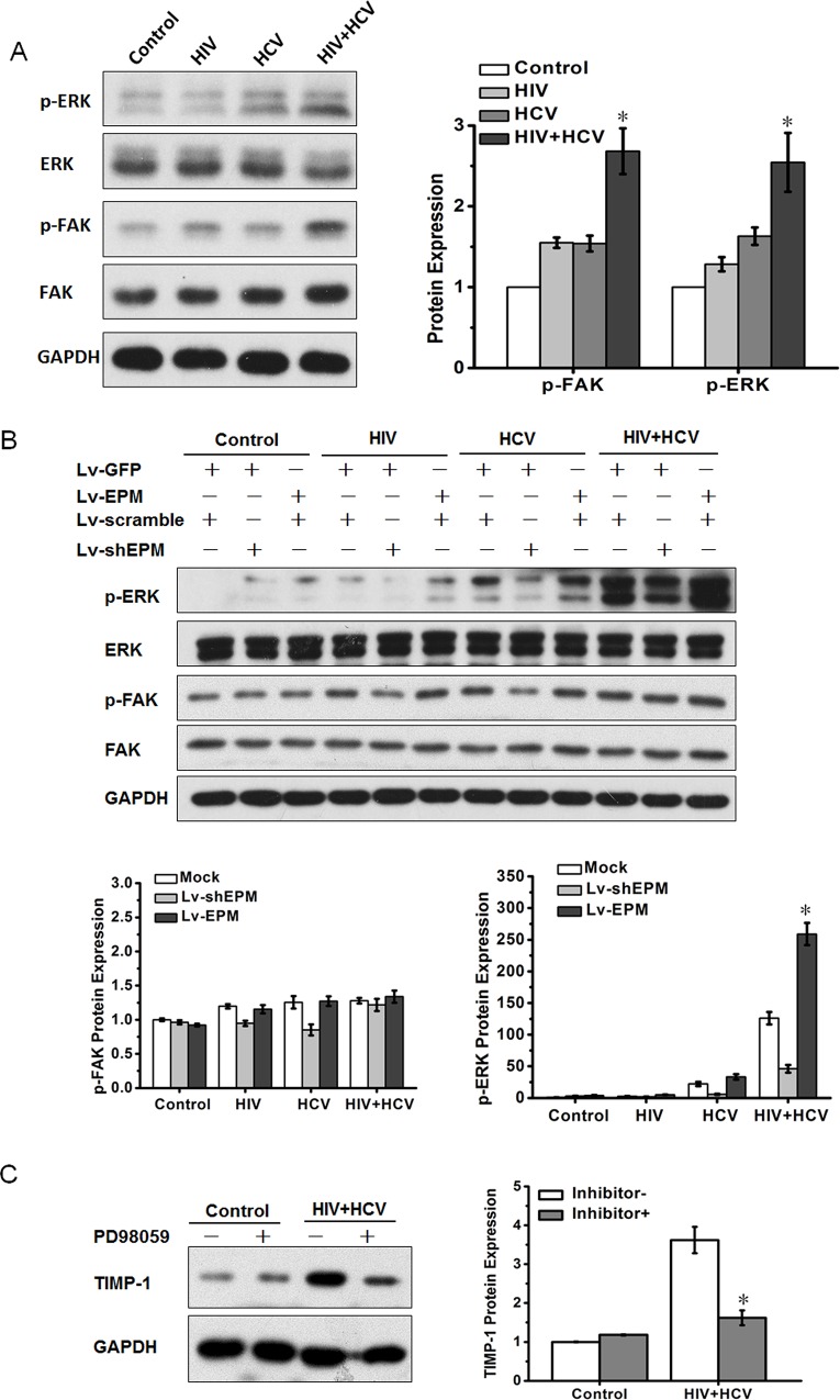
Fig 5. EPM induced TIMP-1 expression via ERK activation.
(A) Western blotting analysis of total protein and phosphorylation levels of FAK and ERK1/2 in LX-2 cells after culture with control medium, HCV, HIV or HIV+HCV, respectively. Bar graphs are shown on the right. (B) Western blotting analysis of total protein and phosphorylation levels of FAK and ERK1/2 in LX-2 cells, which were transfected with EPM knockdown (Lv-shEPM, with Lv-scramble as the control) and EPM overexpression (Lv-EPM, with Lv-GFP as the control), incubated with control medium, HCV (JFH1), inactivated HIV (NL4-3) or HIV and HCV (HIV+HCV). Bar graphs are shown on the right. (C) LX-2 cells were treated with the ERK inhibitor, PD98059 (50 μM) for 24 h, then TIMP-1 expression was assessed using western blotting. Bar graphs are shown on the right.

