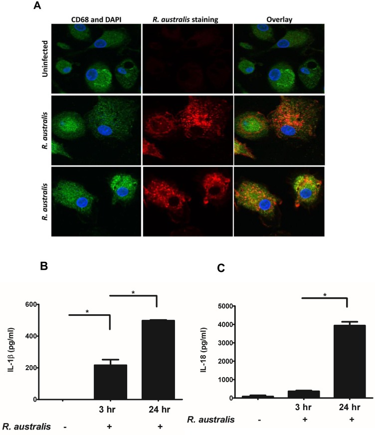
Fig 1. Infection of human PBMC-derived macrophages with R. australis and activation of inflammasome.
Human PBMC-derived macrophages were prepared and infected with R. australis. Cells and culture supernatant were collected at 24 h p.i.. A, Cytosolic rickettsiae were detected by confocal immunofluorescence microscopy after infection with R. australis at an MOI of 5. Images were acquired using 60 × magnification with a water immersion lens. Macrophages are depicted in green, nuclei (DAPI) in blue, and R. australis in red. Secretion levels of IL-1β (B) and IL-18 (C) at 3 h and 24 h p.i. by human PBMC-derived macrophages infected with R. australis at an MOI of 2 were determined by ELISA. Data represent two independent experiments with consistent results. Each experiment included at least 4 replicates. *, p<0.05.

