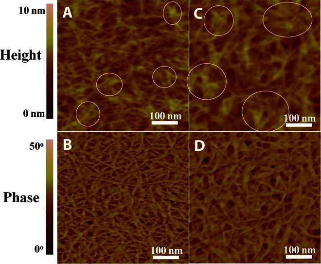
Fig. 6. AFM images of DPPTTT and DPPTTT-NMe4I.

(A to D) AFM height and phase images of the neat DPPTTT thin film (A and B) and the DPPTTT-NMe4I thin film at a molar ratio of 30:1 (C and D). The circles in (C) highlight the formation of more connected and larger fiber aggregates within the DPPTTT-NMe4I thin film in comparison with those marked by circles in (A) for the neat DPPTTT thin film.
