Fig. 2.

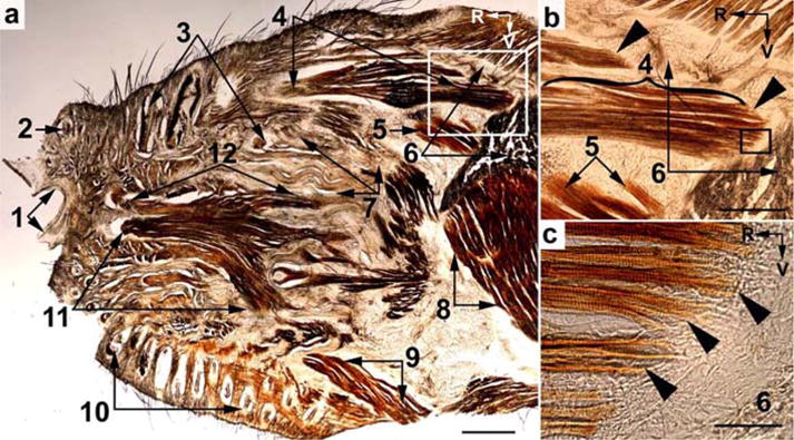
Light microscopy of a tangential slice of the snout of a 2-week-old rat (a). (b) and (c), igher magnification of the boxed areas in (a) and (b), respectively. The slice was stained for CO activity. Arrow heads point at the tapered muscle fibers at the origin of the M. dilator nasi. (1) Nostril; (2) follicle of a nasal vibrissa; (3) follicles of vibrissae in row A; (4) belly of M. dilator nasi; (5) anterior unit of the medial layer of the masseter muscle; (6) zygomatic notch and transversal profile at the base of the processus zygomaticus of the maxilla; (7) subcapsular fibrous mat; (8) anterior part of the masseter muscle; (9) Pars orbicularis oris of the M. buccinatorius; (10) furry buccal pad; (11) and (12) Partes maxillares profunda and superficialis, respectively, of the M. nasolabialis profundus. R, rostral; V, ventral. Scale bars = 1 mm in (a), 0.5 mm in (b), and 0.1 mm (c).
