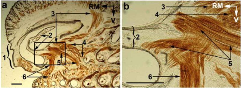
Fig. 4.

Muscle attachments to the cartilages of the nasal sidewall. The boxed area in a is shown at a higher magnification in b. (1) rhinarium; (2) atrioturbinate; (3–5) superficial, pseudointrinsic and posterior slips of the Pars interna respectively; and (6) Pars anterior of the M. nasolabialis profundus. RM, rostromedial; V, ventral. Scale bar: 1 mm. (Sebastian, can you provide another picture of maxillaris profunda of the muscle nasolabialis profundus.)The one above was taken from one of your prior papers)
