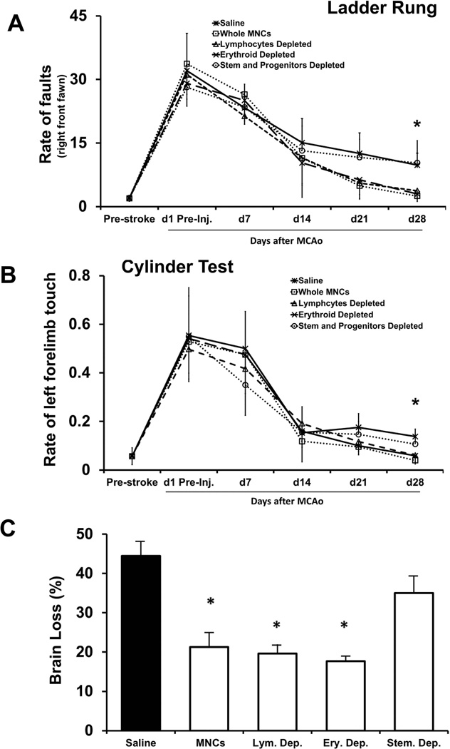
Fig 1. Functional improvement in mice treated with whole MNCs and MNCs depleted of specific sub-populations of cells via IV at 24 hrs after stroke.
Mice were assigned to 6 treatment groups: 1) saline; 2) whole MNCs; 3) MNCs with Lymphoid cell depletion; 4) MNCs with Myeloid cell depletion; 5) MNCs with Erythroid lineage cell depletion; 6) MNCs with stem cell and progenitor cell depletion. Line diagrams illustrating evaluation on the Cylinder (A) and Ladder run (B) tests up to 28 days after stroke. Data are mean ± SD; N= 12 per group initially but only 6 survived to the end of observation and all mice with myeloid cell depleted MNCs died within 9 days after stroke. *P < 0.05, compared with saline controls. The bar graph (C) exhibiting significantly reduced cerebral atrophy among the same experimental groups at 28 days after stroke. Data are mean ± SD. N= 6 per group. *P < 0.05 compared with saline controls.

