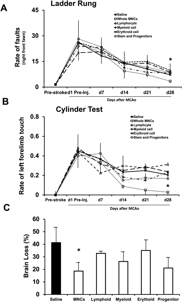
Fig 4. Functional improvement in mice treated with whole MNCs or a single cell lineage component within MNCs via IV delivery at 24 hrs after stroke.
Mice were randomly assigned to 6 treatment groups after MCAo and autologous MNCs positive selection:1) saline; 2) whole MNCs; 3) Lymphoid cell within MNCs; 4) Myeloid cell within MNCs; 5) Erythroid lineage cell within MNCs; 6) Stem cell and progenitor cell within MNCs. Line diagrams illustrating the evaluation on the Ladder run (A) and Cylinder (B) tests up to 28 days after stroke. The bar graph (C) exhibiting the cerebral atrophy among the same experimental groups at 28 days after stroke. Data are mean ± SD. N= 12 per group initially and N= 6–8 at the end of experiment per group. *P < 0.05 compared with saline controls.

