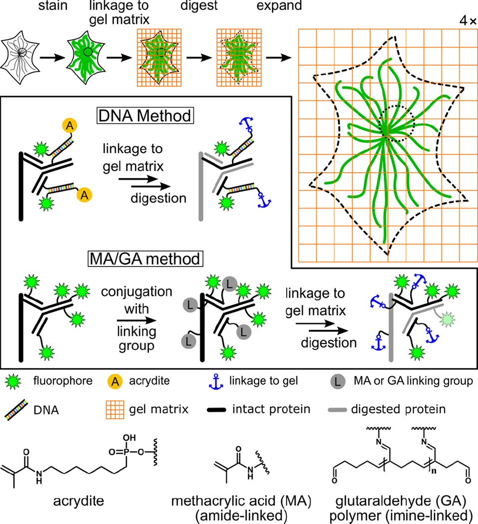
Figure 1.
Schematic illustration of expansion microscopy and label retention strategies. The boxed region highlights the difference between the original DNA method1 and the post-stain linker-group functionalization method (“MA/GA method”) presented in this work. In the DNA method, the specimen is immunostained with a custom-prepared antibody bearing doubly-modified DNA linked to a fluorophore and an acrydite moiety (A). In contrast, with the MA/GA method, methacrylic acid N-hydroxy succinimidyl ester (MA-NHS) or glutaraldehyde (GA) are used to label the entire sample with polymer-linking groups after conventional immunostaining with fluorophore-labeled antibodies (only secondary antibodies are shown). For both methods, the next steps are gelation, digestion with a protease, and expansion through dialysis into deionized water. The acrydite (A), MA, and GA groups allow formation of a linkage to the hydrogel. Dyes are retained through a connection to antibody fragments that also contain a linkage to the gel. Fluorescent proteins are also retained using the MA/GA method through a similar method but are not shown here for the sake of clarity.

