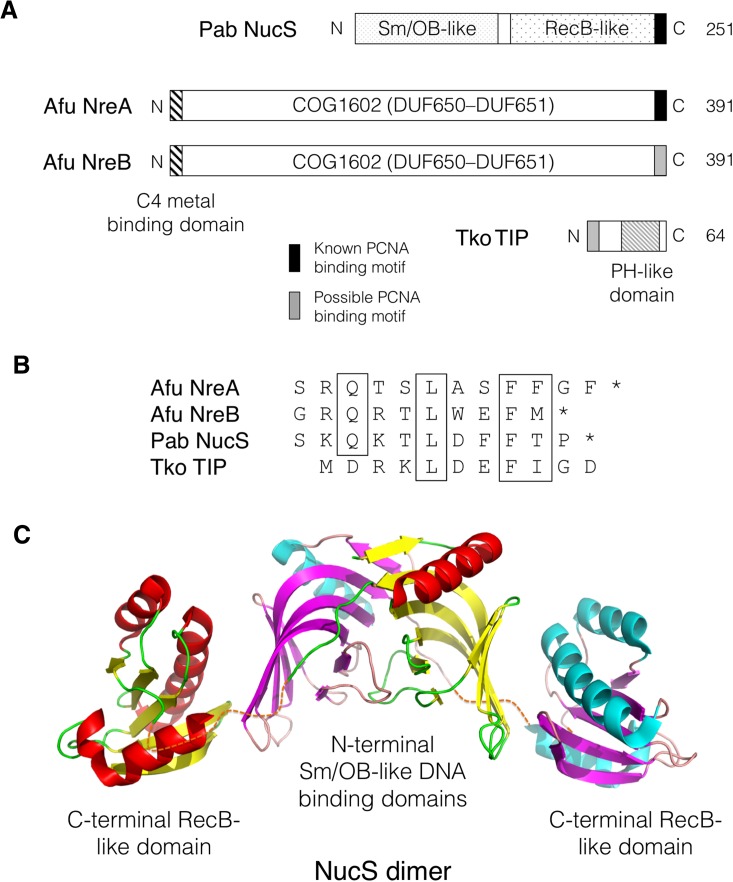
Fig. 2.
a Schematic representation of the P. abyssi NucS, A. fulgidus NreA and NreB and T. kodakarensis TIP proteins highlighting conserved domains. b Known (NucS, NreA) or possible (NreB, TIP) PIP motifs in the four proteins with conserved residues boxed (see Supplementary information, Figure S1, for full TIP alignment). c Three-dimensional structure of residues 1–233 of the P. abyssi NucS protein dimer (PDB code 2VLD). Secondary structure is coloured in red/yellow/green (monomer 1) and cyan/magenta/pink (monomer 2). Broken lines indicate the interdomain linker region (residues 115–125) not seen in crystal structure. The C-terminal region (residues 234–251) that includes the PIP motif is also absent from the structure. See text for details and references

