. 2016 Jun 24;19(2):191–198. doi: 10.4048/jbc.2016.19.2.191

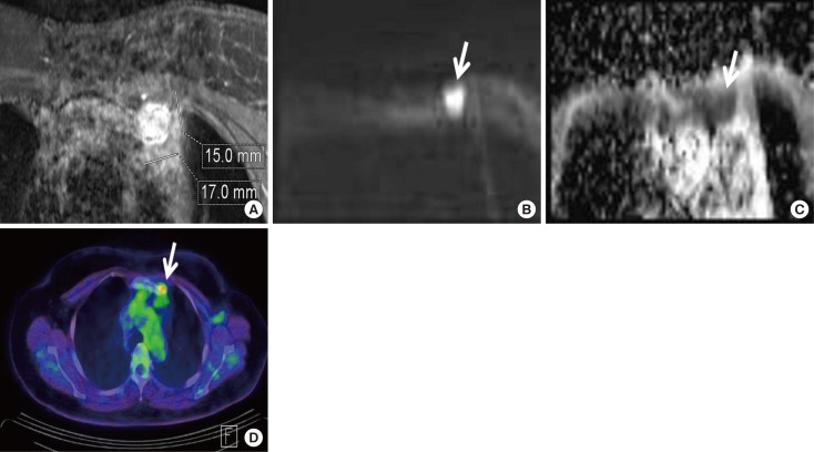
Figure 1. A 41-year-old woman with invasive ductal carcinoma in the left breast and malignant lymph node confirmed by core needle biopsy. (A) Axial fat suppressed contrast enhanced T1-weighted image reveals an internal mammary lymph node in the first intercostal space with short axis of 15 mm, long axis of 17 mm and loss of fatty hilum. (B) Diffusion weighted imaging shows a high-signal-intensity of lymph node (arrow). (C) Apparent diffusion coefficient map shows a low-signal-intensity (arrow). (D) Positron emission tomography computed tomography reveals presence of focal fluorodeoxyglucose uptake in the same location of left internal mammary lymph node with a maximum standardized uptake value of 5.6 (arrow).

Figure 1