. 2016 Jun 24;19(2):191–198. doi: 10.4048/jbc.2016.19.2.191

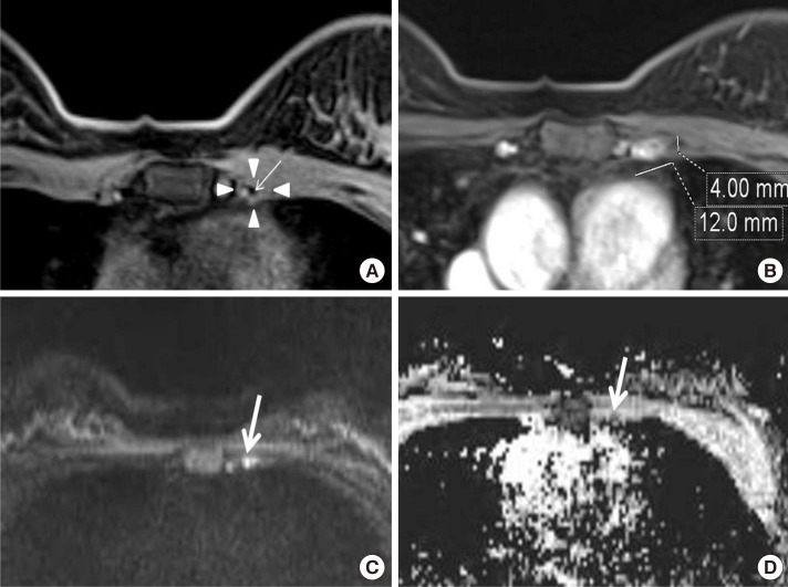
Figure 3. A 41-year-old woman with invasive ductal carcinoma in the left breast and benign lymph node confirmed by fine-needle aspiration and biopsy. (A) Axial fat suppressed T1-weighted image reveals an internal mammary lymph node (arrowheads) in the second intercostal space with preserved fatty hilum showing low-signal-intensity (arrow). (B) On axial fat suppressed contrast enhanced T1-weighted image, the size of lymph node is 4 mm (short axis) and 12 mm (long axis). (C) Diffusion weighted imaging shows a high-signal-intensity of lymph node (arrow). (D) Apparent diffusion coefficient map shows a iso-signal-intensity (arrow).

Figure 3