Figure 1.
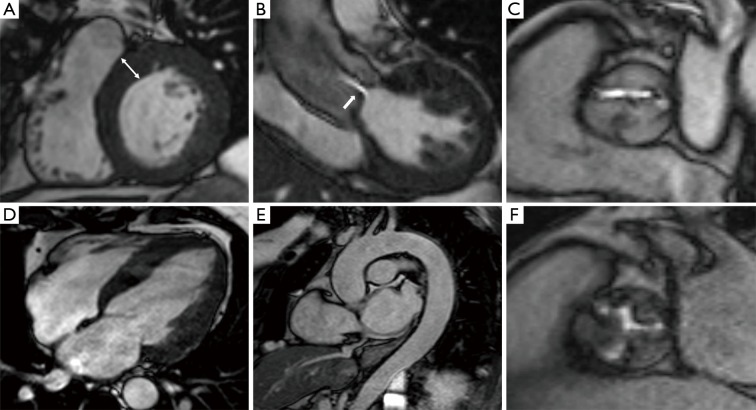
Cardiovascular magnetic resonance (CMR) cine imaging demonstrating anatomical and functional information. (A) Short-axis of left ventricle at the basal level in diastole indicating mild concentric hypertrophy (white arrow, 14 mm); (B) coronal left ventricular outflow tract (LVOT) view acquired through-plane showing the aortic valve leaflet tips and restricted leaflet motility and resultant high velocity jet (white arrow); (C) cine imaging of a bicuspid aortic valve orifice in systole with A-P closure line, this view permits direct planimetry of valve area in addition to morphological assessment; (D) 4-chamber view allowing visual assessment of ventricular, mitral and tricuspid function and atrial size; (E) sagittal-oblique view of aorta throughout its entire thoracic course; (F) cine imaging of heavily stenosed trileaflet aortic valve.

