Figure 1.

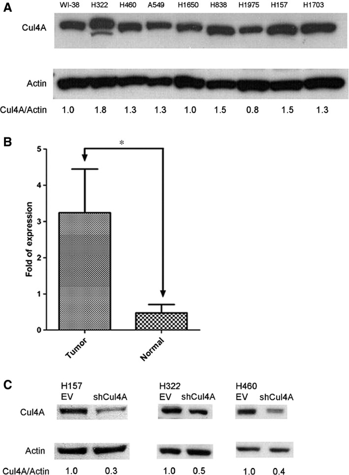
(A) Expression of Cul4A in normal lung and lung cancer cell lines. Cul4A in normal lung (WI‐38) and lung cancer (H322, H460, A549, H1650, H838, H1975, H157 and H1703) cell lines. Internal control: β‐Actin. Density of Cul4A bands was quantified and normalized to β‐Actin using WI‐38 cell line as a normal control. (B) Expression of Cul4A mRNA in NSCLC tumour and paired normal lung tissue samples. Bars represent the average of fold of expression ± standard deviation (n = 32). ‘*’ denotes P < 0.0001, t‐test. (C) Knockdown of Cul4A expression by shRNA. Density of Cul4A bands was quantified and normalized to β‐Actin using empty vector‐transfected cell lines as normal controls. EV: empty vector; shRNA: Cul4A shRNA.
