Figure 3.

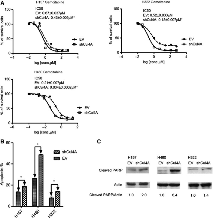
(A) IC 50 values for gemcitabine in Cul4A shRNA transfected H157, H460, and H322 lung cancer cell lines. Data points represent the average of IC 50 value± standard deviation of gemcitabine in triplet experiments. EV: empty vector; Cul4A: Cul4A shRNA; ‘*’ denotes P < 0.05, t‐test. (B) Apoptosis assay (Annexin‐V FITC) in lung cancer cell lines after treated with 5 μM gemcitabine for 72 hrs. Percentage of apoptotic cells were shown as bar ± standard deviation in triplet experiments. EV: empty vector; Cul4A: Cul4A shRNA; ‘*’ denotes P < 0.05, t‐test. (C) Western blot analysis for cleaved PARP. Lung cancer cell lines were treated with 5 μM gemcitabine for 24 hrs and were harvested. Total cell lysate was used for Western blot analysis with cleaved PARP antibody (Cell Signaling). Density of cleaved PARP bands were quantified and normalized to β‐Actin using empty vector transfected cell lines as normal controls. EV: empty vector; Cul4A: Cul4A shRNA.
