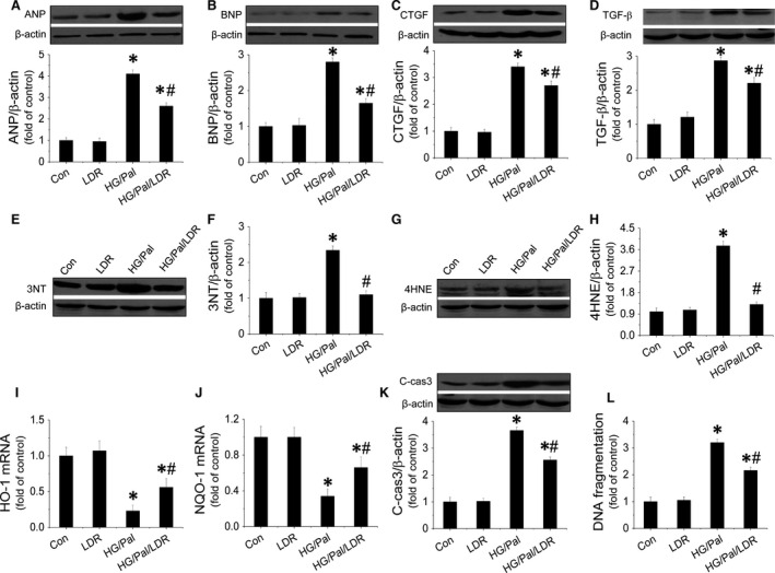
Figure 4.

LDR prevented hypertrophy, fibrosis, oxidative stress and apoptosis in HG/Pal‐treated primary cardiomyocytes. Primary cardiomyocytes from adult mice were isolated and treated with high glucose (33 mmol/l) for 24 hrs associated with palmitate (62.5 μmol/l) during the last 15 hrs. Cells were exposed to LDR at 25 mGy every 6 hrs initiated just before high‐glucose treatment. Western blot assay was applied to identify cardiomyocyte hypertrophy by measuring expressions of ANP (A) and BNP (B); fibrosis in cardiomyocytes by measuring expressions of CTGF (C) and TGF‐β (D); oxidative stress in cardiomyocytes by examining the expression 3‐NT (E and F), 4‐HNE (G and H), HO‐1 mRNA (I) and NOQ‐1 mRNA (J); and cell apoptosis by quantifying expression of cleaved‐caspase‐3 (K) and DNA fragmentation (L). Data are presented as means ± S.D., n = 8/group. *P < 0.05 versus the Con group; # P < 0.05 versus the DM group.
