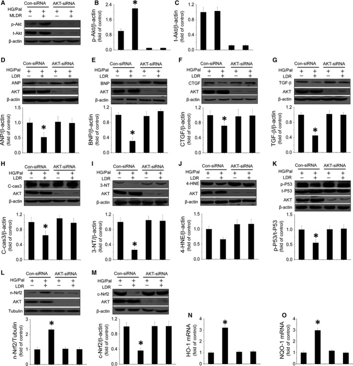
Figure 5.

Akt mediates LDR‐induced antihypertrophic and antifibrotic effects against HG/Pal associated with suppression of P53‐induced apoptosis and improvement of Nrf2 translocation and transcriptional function. Primary cardiomyocytes were transfected with either negative control sense siRNA or mouse Akt antisense siRNA. Western blot was used to measure Akt phosphorylation (A and B) and expression (A and C). Expression of ANP (D) and BNP (E) as markers of hypertrophy; CTGF (F) and TGF‐β (G) as markers of fibrosis were measured by Western blot assay. Expression of cleaved‐caspase3 (H) and 3‐NT (I), 4‐HNE (J) reflecting apoptosis or oxidative stress were quantified. To measure mediators of both apoptotic and oxidative pathways induced by HG/Pal treatment, P53 activity (K) and Nrf2 translocation (L and M) and transcriptional function (N and O) were evaluated by Western blot. Data are presented as means ± S.D., n = 8/group. *P < 0.05 versus the Con group; # P < 0.05 versus the DM group.
