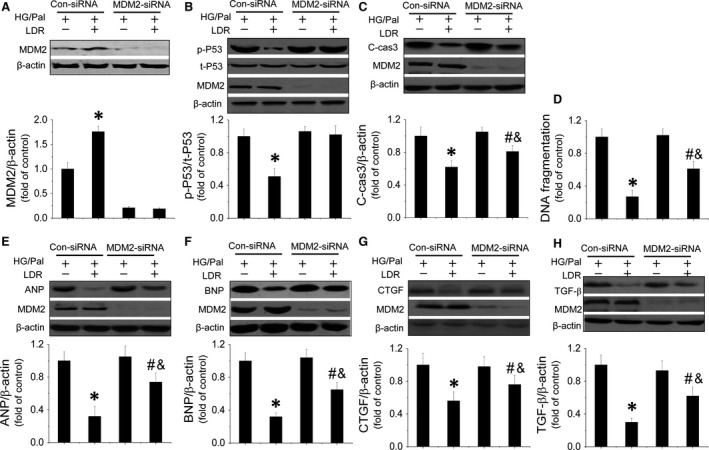
Figure 6.

MDM2/P53‐mediated anti‐apoptotic pathway is involved in LDR‐induced cardiac protection in vitro. TO evaluate the relationship between MDM2/P53‐mediated anti‐apoptotic effects and LDR‐induced cardiac protection in vitro against HG/Pal. Primary cardiomyocytes were transfected with either negative control sense siRNA or mouse MDM2 antisense siRNA using Lipofectamine TM 2000 transfection reagent for 48 hrs. Transfection was followed by treatment with HG/Pal with/without exposure to LDR at 25 mGy. Western blot was used quantify P53‐mediated apoptosis by measuring MDM2 expression (A) P53 phosphorylation and cleaved‐caspase3 expression (B and C). Meanwhile another apoptotic marker, DNA fragmentation, was measured by ELISA (D). Additionally, expression of hypetrophic markers, ANP (E) and BNP (F) and fibrotic markers, CTGF (G) and TGF‐β (H) were measured by Western blot. Data are presented as means ± S.D., n = 8/group. *P < 0.05 versus the Con group; # P < 0.05 versus the DM group.
