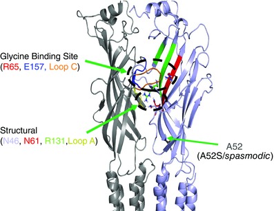
Figure 9. Structural model of the GlyR .

Model showing a side‐view of the GlyRα1 subunit interface highlighting the important structural residues, N46, N61 and R131, all residing in the same plane as loop A and positioned below the orthosteric glycine binding site, involving residues: R65, E157 and loop C.
