
Figure 2. The structure of repair Schwann cells .
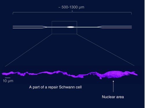
A repair Schwann cell in the distal stump (4‐week transected tibial nerve without re‐innervation), as shown by serial block face scanning electron microscopy. Only a part of the cell is shown, as indicated by the box superimposed on a schematic diagram of a repair cell (R. Mirsky, K. R. Jessen H. Armer and P. Munro, unpublished).