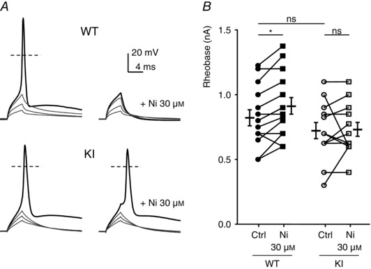
Figure 4. Action potential (AP) properties in DRG neurons from WT and KI mice .

A, typical traces of evoked APs. Depolarization currents of increasing amplitude were injected to trigger an AP in the absence (left) or presence of 1 μm zinc (right) in D‐hair neurons from WT (upper panels) and KI (lower panels) mice. The resting potential of these neurons was maintained at −70 mV. B, threshold current for an AP generation (Rheobase) in control condition and with 30 μm nickel for neurons from WT mice (left) and KI mice (right). Statistical analysis was performed with repeated‐measures two‐way ANOVA (WT Ctrl vs. WT Ni 30 μm, *P < 0.05; WT Ctrl vs. KI Ctrl, P = 0.52; WT Ni 30 μm vs. KI Ni 30 μm, P = 0.08; KI Ctrl vs. KI Ni 30 μm, P = 0.95).
