
Figure 7. NeuroD2 regulates AHP through SK2 .
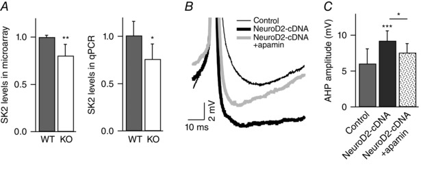
A, SK2 mRNA levels are decreased in NeuroD2 KO tissue as measured by microarray (n = 7 for both WT and KO) and qPCR (n = 5 experiments with each in triplicate for both WT and KO). Data are from 14 DIV cultured cortical neurons (*P < 0.05; **P < 0.01). B, representative traces of AHP after the first AP (in 8 Hz AP trains) in control, NeuroD2‐cDNA, and NeuroD2‐cDNA + apamin. C, summary of AHP measurements after the first AP in control, NeuroD2‐cDNA and NeuroD2‐cDNA + apamin (*P < 0.05, ***P < 0.001).