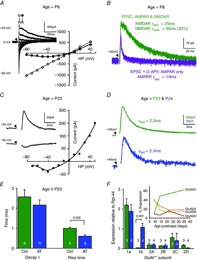
Figure 7. LSO NMDAR‐mediated EPSC decay time accelerates during development .

A, evoked EPSC I–V relationship from a pre‐hearing (P8) LSO principal neuron (in the presence of 10 μm bicuculline and 1 μm strychnine). Open circles indicate the latency at which the I–V of the fast AMPAR‐EPSC is measured, closed circles show the latency for the I–V of the NMDAR‐EPSC. Inset: superimposed average EPSC traces for each holding potential (HP) from −80 to +40 mV. Throughout, the stimulus is indicated by a triangle, with artifacts being removed for clarity. B, in a Pre‐hearing mouse (P8) an LSO neuron held at a potential of +40 mV shows the evoked EPSC before (green trace) and after perfusion of 20 μm d‐AP5 in the same cell. The fast AMPAR‐mediated response remains in the presence of AP5 (purple trace) and confirms that the slow EPSC is mediated by NMDAR. The EPSC time‐constants are indicated by the respective trace. C, in a mature mouse (P23) the EPSC from an LSO neuron is recorded in the presence of 10 μm bicuculline, 1 μm strychnine and 10 μm NBQX to isolate the NMDAR component. Inset: average traces from HP of +50 and −50 mV. Note the voltage‐dependence of the EPSC and the fast decay time‐course of this NMDAR‐mediated EPSC. D, NMDAR‐EPSCs from Control (green) and AT‐exposed (blue) mice (>P23) show similar fast decay time‐courses at +40 mV HP. E, bar chart showing mean NMDAR‐EPSC decay τ and rise time for control and AT‐exposed mice, respectively. F, relative expression of GluN1 and GluN2 subunit mRNA in the LSO from control (green) and following AT exposure (blue). GluN1b mRNA at P35 was significantly increased following AT. AT in mature mice increased GluN1b mRNA and decreased the NMDAR‐EPSC rise time, but had no impact on decay time‐course. Inset: developmental profile of GluN2 subunit mRNA expression shows GluN2C mRNA levels increase and dominate over maturation. Statistical comparison was performed using a t test in E and one‐way ANOVA in F with P values as indicated.
