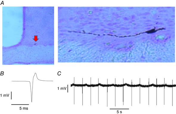
Figure 1. Juxtacellularly labelled neurone in the SCN .

A, juxtacellularly labelled neurone (arrowed) in the SCN, shown at higher magnification on the right. B, average action potential from the labelled neurone (average of 1000 spikes). The cell was recorded during the subjective day. C, extract of voltage trace showing individual spikes. Note the regularity of discharge. OC, optic chiasm; 3V, third ventricle.
