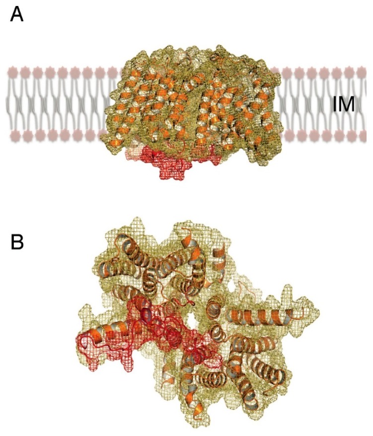
Figure 2.
The crystal structure of MraY from Aquifex aeolicus (PDB 4J72) reveals (A) a dimer displaying 10 TM helices per monomer, whose N- and C-termini face the periplasm. (B) A view from the cytoplasmic side indicates a tunnel formed in the monomer-monomer interaction region, buttressed by cytoplasmic loops (in red) that could interact with substrate, partner proteins, or both.

