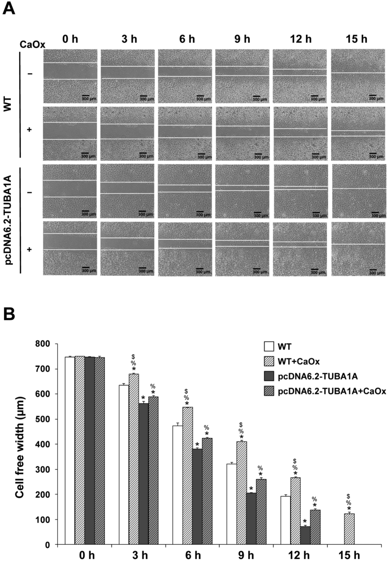
Figure 6. Effect of α-tubulin overexpression on tissue repair.
Evaluation of tissue repair was done by using a scratch assay. (A) Representative images of cell-free width used for quantitative analysis (original magnification power was 40X). (B) Quantitative and statistical analyses of cell-free width at 0–15 h. The data are reported as mean ± SEM (n = 3 independent experiments). *p < 0.05 vs. WT; **p < 0.01 vs. WT; ##p < 0.01 vs. WT + CaOx; %p < 0.05 vs. pcDNA6.2-TUBA1A; $p < 0.05 vs. pcDNA6.2-TUBA1A + CaOx.

