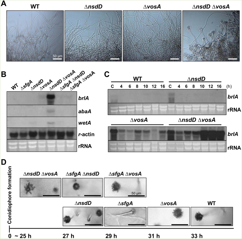
Figure 2. Accelerated conidiation by the lack of vosA and nsdD.
(A) Photographs of WT (FGSC4), ΔnsdD (TNJ108), ΔvosA (THS15) and ΔnsdD ΔvosA (TMK11) hyphae at 16 h in liquid MMG. (Bar = 50 μm). (B) Levels of brlA, abaA, and wetA mRNA in designated strain grown in liquid submerged culture for 16 h. The γ-actin gene was used as a control. (C) Levels of brlA mRNA in WT, Δnsd, ΔvosA and ΔnsdD ΔvosA strains in liquid submerged culture up to 16 h. C = conidia. (D) Time needed for the formation of the first conidiophore in single colonies of WT (FGSC4), ΔnsdD (TNJ108), ΔvosA (THS15), ΔsfgA (TNJ57), ΔsfgA ΔnsdD (TMK5), ΔsfgA ΔvosA (TMK10), and ΔnsdD ΔvosA (TMK11) on solid MMG. Photographs were taken at indicated time when the first conidiophore was visible. Numbers indicate the incubation time (h) after streak on solid MMG.

