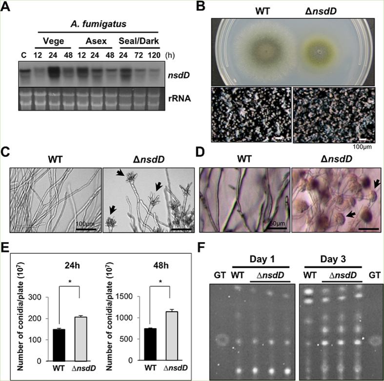
Figure 4. Characterization of nsdD in A. fumigatus.
(A) Levels of nsdD mRNA during the life cycle of A. fumigatus WT (AFU293). C = conidia. See Fig. 3B legend for the times. Equal loading of total RNA was confirmed by ethidium bromide staining of rRNA. (B) Phenotypes of A. fumigatus WT and ΔnsdD (FNJ19) strain point inoculated on solid MMG with 0.1% YE and incubated at 37 °C for 3 days. Close-up views (lower panel) of the center of the colonies are shown (Bar = 100 μm). (C) Cells of A. fumigatus WT and ΔnsdD strains grown in liquid submerged culture for 19 h (Bar = 100 μm). Note abundant formation of aberrant conidiophores in the mutant. Conidiophore is marked by arrowhead. (D) Agar-embedded cells of A. fumigatus WT and ΔnsdD strains grown on solid MMG for 27 h (Bar = 50 μm). A high number of conidiophores was evident. (E) Quantitative analysis of conidiation: 105 spores of WT and ΔnsdD strains were spread on solid MMG with 0.1% YE, incubated for 24 and 48 h, and the conidia numbers per plate were counted in triplicates (*P < 0.001). (F) Thin-layer chromatogram of CHCl3 extracts of A. fumigatus WT and ΔnsdD strains grown in liquid MMG with 0.5% YE for 1 and 3 days (stationary culture). Gliotoxin standard (GT) is shown.

