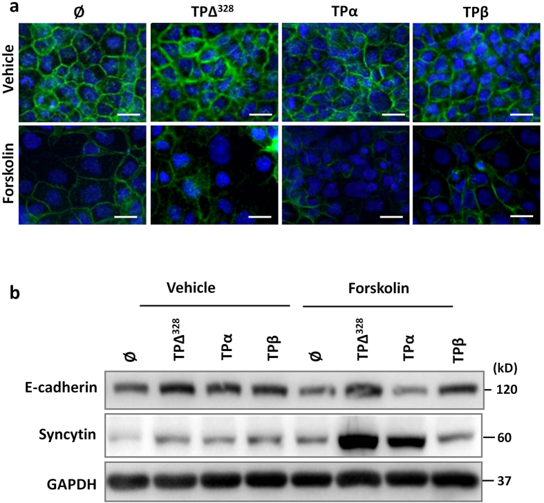
Figure 4. TPβ slows and TPα enhances the rate of syncytialisation of trophoblast cells.
(a) Transfected BeWo cells were incubated for 72 hours with 200 nM I-BOP (vehicle) or 200 nM I-BOP and 100 μM forskolin (forskolin) to induce syncytialisation. Membrane fusion was monitored by staining for E-cadherin (green), and cell number demonstrated with DAPI (blue). Cells were imaged at 400x magnification, scale bar: 20 μm. (b) Similarly cultured, transfected BeWo lines were assessed for changes in E-cadherin and syncytin expression via immunoblotting. GAPDH was used to control for protein loading and protein bands were quantified using densitometry. Data are representative of 3 independent experiments.

