Figure 1.
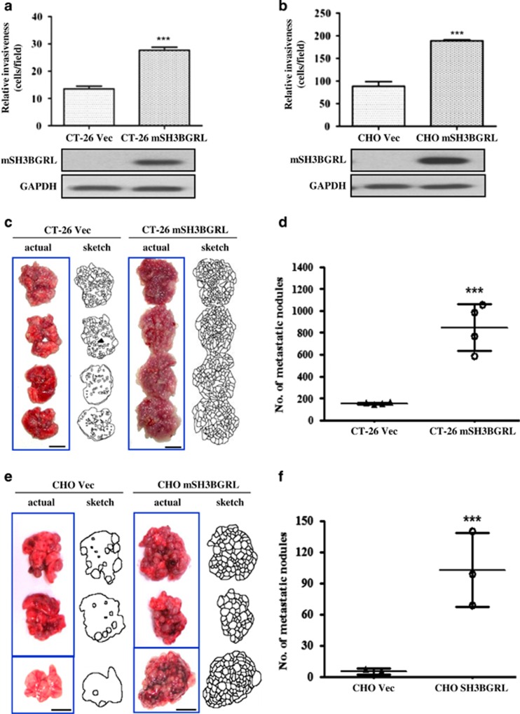
mSH3BGRL enhances cell invasiveness in vitro and drives metastasis in vivo. (a) Matrigel invasion assay using CT-26 cells stably overexpressing vector (CT-26 Vec) or GFP-SH3BGRL (CT-26 mSH3BGRL). After 36 h incubation, the number of invaded cells were counted (mean±s.d., n=3, ***P<0.001). The accompanying immunoblot shows the expression of exogenous SH3BGRL, with glyceraldehyde 3-phosphate dehydrogenase (GAPDH) as a loading control. (b) Similar experiment as in (a), but using CHO cells stably expressing vector (CHO Vec) or RFP-SH3BGRL (CHO mSH3BGRL) (mean±s.d., n=3, ***P<0.001). (c) In total, 1 × 10⁶ CT-26 Vec or CT-26 SH3BGRL cells were injected intravenously into the tail vein of nude mice. After 28 days, mice were killed and their lungs were photographed. (d) Scoring for metastatic tumor nodules in (c) (mean±s.d., n=4, ***P<0.001). (e) In total, 1 × 10⁶ CHO Vec or CHO mSH3BGRL cells were injected and analyzed after 28 days injection. (f) Scoring for metastatic tumor nodules as in (e) (mean±s.d., n=3, ***P<0.001).

