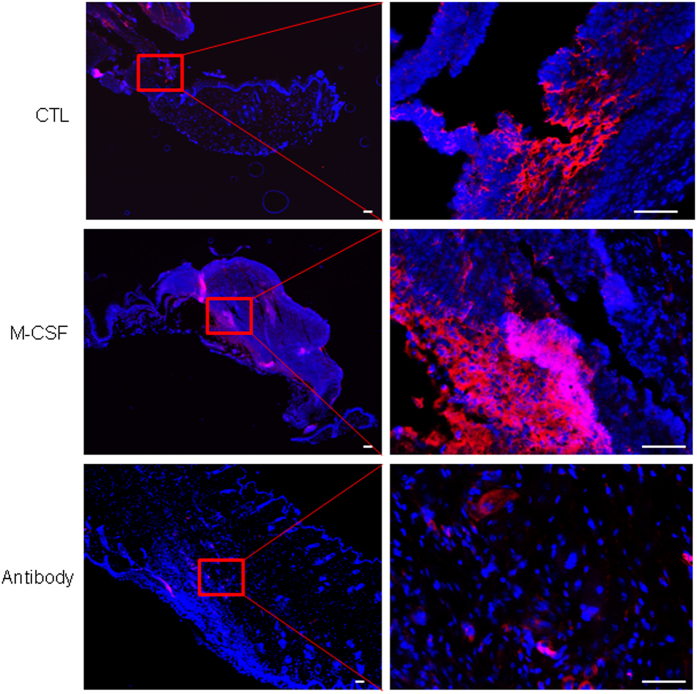
Figure 4. Injury-induced SSEA-1 positive cells were regulated by topical application of M-CSF or M-CSF neutralizing antibody.
Skin wounds in mice were treated by either PBS control (top panels) or topical application of recombinant M-CSF protein (middle panels) or topical application of M-CSF neutralizing antibody for 6 days. This figure showed a representative skin section from 6 wounds in each group, stained with SSEA-1 (red color). DAPI (blue) was used for cell nucleus staining. Scale bars, 100 μm.

