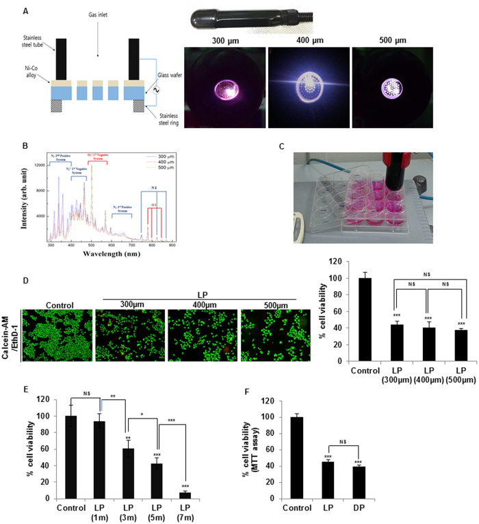
Figure 1. Preparation of liquid plasma (LP) using a microplasma system and the effects on cancer cells.
(A) A photo of the hand-held-type system that is a microplasma jet device, and photographs of the air microplasma jet generated at atmospheric pressure by means of a nozzle with pores 300–500 μm in diameter. (B) Optical emission spectra of air microplasma jets during discharge in the wavelength range 280–920 nm with the end diameters of the collimated plasma jets from 300 to 500 μm. (C) Preparation of water plasma via exposure of a medium to plasma located at a ~2-cm distance. Media (DMEM or RPMI 1640) supplemented with 1% fetal bovine serum (FBS) were added to wells of a 12-well-plate (1 mL per well), and then exposed to the plasma jet for indicated periods. (D) Viability of HeLa cells was assessed by the live/dead assay after treatment of the cells with 5 min LP generated by means of 300-, 400-, or 500-μm collimated plasma jets. Representative images of calcein-AM/EthD-1 - stained cells under a fluorescence microscope are shown. The right graph shows static analysis of viability of LP-treated HeLa cells compared to untreated (control) cells. (E) HeLa cells were treated with liquid plasma generated by air plasma spraying above the media surface for the indicated times (1, 3, 5, and 7 min), and Live/dead assay was performed as described in Materials and Methods. (F) Comparison of HeLa cell viability by the MTT assay after 24 h of treatment with liquid or direct plasma. NS, not significant; *P ≤ 0.05, **P ≤ 0.01, ***P ≤ 0.001.

