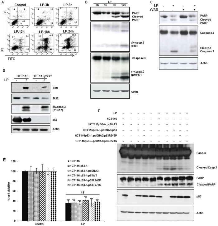
Figure 4. Liquid plasma (LP) induces apoptotic cell death in a p53-independent manner.
HeLa cells were treated with LP for the indicated periods. (A) Cell death was analyzed by fluorescence-activated cell sorting (FACS) following Annexin V and propidium iodide (PI) staining. (B) Activation of the caspase cascade after LP treatment. Immunoblotting was performed with antibodies against cleaved caspase 8 and cleaved caspase 3, PARP, and actin (cropped, full-length images are shown in Supplementary Fig. S4A). (C) A caspase inhibitor attenuates LP-induced apoptosis. HeLa cells were treated with z-VAD for 1 h before LP treatment. After 24 h, proteins in cell lysates were separated by sodium dodecyl sulfate polyacrylamide gel electrophoresis (SDS-PAGE) and immunoblotted with antibodies against cleaved caspase 3, PARP, and actin (cropped, full-length images are shown in Supplementary Fig. S4B). (D) HCT116 wild-type, and HCT116 p53−/− cells were treated with LP and harvested after 24 h. BH3 protein (proapoptotic Bim) was activated, and prosurvival Bcl-2 was downregulated in response to LP, in a p53-independent manner. Immunoblotting was performed with antibodies against Bim, Bcl-2, caspase 3, p53, and actin (cropped, full-length images are shown in Supplementary Fig. S4C). (E, F) Cell viability of HCT116 wild-type, HCT116 p53−/−, and HCT116 p53−/− cells with reconstituted p53 (wild type or mutants) was measured by the live/dead assay (E) and cellular apoptosis was evaluated by western blot of cleaved caspase 3 and PARP (F) after 24 h of LP treatment (cropped, full-length images are shown in Supplementary Fig. S4D). Expression of endogenous or exogenous p53 in these cells was confirmed by western blotting and the amount of actin protein was served as a loading control. NS, not significant; *P ≤ 0.05, **P ≤ 0.01, ***P ≤ 0.001.

