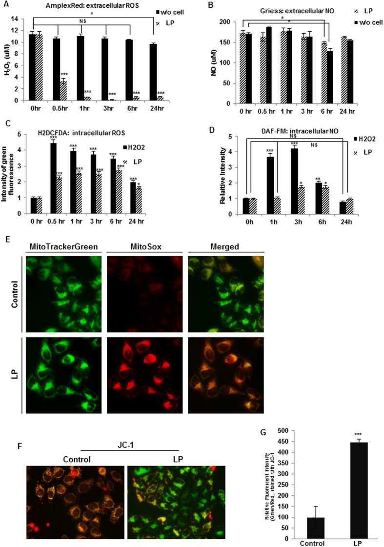
Figure 5. Liquid plasma (LP) generated reactive oxygen and nitrogen species (ROS and RNS), and induced mitochondrial ROS accumulation, resulting in reduced mitochondrial membrane potential (MMP).
Extra- and intracellular ROS and RNS were quantified by the AmplexRed assay (extracellular H2O2, (A)), the Griess assay (extracellular NO, (B)), 5,6-carboxy-2′,7′-dichlorofluorescein diacetate (H2DCF-DA) assay (intracellular ROS, (C)), and DAF-FM assay (intracellular NO, (D)). (E) Mitochondrial ROS accumulation was assessed by staining the cells with MitoTracker Green and MitoSox Red. (F,G) MMP of HeLa cells was assessed under a fluorescence microscope (F) or measured by fluorescence-activated cell sorting (FACS) (G) following 5,5′,6,6′-tetrachloro-1,1′,3,3′-tetraethyl-benzimidazolylcarbocyanine chloride (JC-1) staining. NS, not significant; *P ≤ 0.05, **P ≤ 0.01, ***P ≤ 0.001.

